
The Brunei Cancer Centre
Nuclear Medicine
Overview
Nuclear Medicine
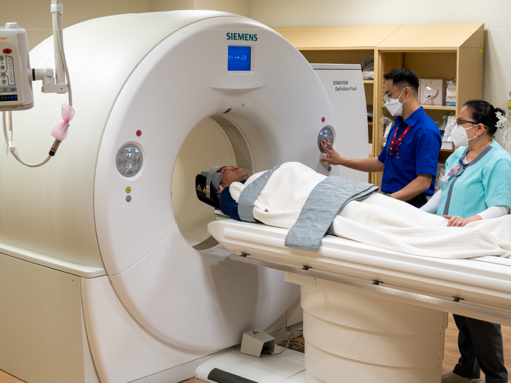
Precision diagnosis and targeted therapy using radiopharmaceuticals and advanced imaging technology. Our state-of-the-art nuclear medicine facilities combine PET-CT, SPECT-CT and specialised cardiology imaging to detect cancer at earlier stages, assess treatment response and guide clinical decision-making. Working alongside our radiation and medical oncology teams, we deliver comprehensive care anchored in molecular medicine. From diagnosis to therapy monitoring, our expert team brings detailed functional and anatomical insight to every case.
What is Nuclear Medicine?
Nuclear medicine is a branch of medicine where radioactive substances, known as radiopharmaceuticals, are used to diagnose and treat both cancerous and non-cancerous conditions through imaging and targeted therapy. These radiopharmaceuticals are absorbed by specific organs or disease sites and emit radiation to create images or provide treatment.
This approach enables physicians to capture detailed images of the body’s internal structures and functions, resulting in accurate diagnoses and enhanced patient outcomes. By combining functional and anatomical information, nuclear medicine provides a comprehensive view of disease activity that conventional imaging alone cannot achieve.
Our Department
The department is licensed by the Safety Health and Environment National Authority (SHENA) and complies with the standards set by the International Atomic Energy Agency (IAEA). We feature a fully equipped nuclear medicine facility with state-of-the-art imaging suites and treatment capabilities.
Regulatory Compliance
Licensed by SHENA and fully compliant with International Atomic Energy Agency (IAEA) standards, ensuring the highest levels of safety and quality in all diagnostic and therapeutic procedures.
Services and Treatments
PET-CT Suite
Positron Emission Tomography – Computed Tomography (PET-CT)
Comprehensive functional and anatomical imaging with Siemens Biograph mCT integrated 64-slice CT.
The PET-CT Suite features a dedicated dose dispensing station equipped with a dose calibrator and a PET tracer injection area monitored by closed-circuit television surveillance. At the core of the suite is the Siemens Biograph mCT, integrated with a 64-slice CT scanner to deliver precise, high-quality PET-CT imaging.
During a PET-CT scan, a small dose of radiopharmaceutical is injected into a vein. The radiopharmaceutical circulates throughout the body and is taken up by targeted cells, providing functional and physiological information. The CT component uses X-ray radiation to produce detailed anatomical images of the body. Together, these two imaging techniques provide comprehensive diagnostic information.
By combining PET and CT data, a detailed hybrid image is produced, allowing accurate localisation and assessment of abnormal uptake within the body. This enables clinicians to evaluate both anatomical structures and physiological activity in a single examination.
Duration
3 to 4 hours from arrival to discharge
Technology
Siemens Biograph mCT + 64-slice CT
Application
Oncology, cardiology, neurology
SPECT-CT Suite
Single Photon Emission Computed Tomography – Computed Tomography (SPECT-CT)
The SPECT-CT Suite features a dedicated radiopharmaceutical injection area and a specialised radio-aerosol inhalation room. Central to the suite is the Siemens Symbia TruePoint SPECT integrated with a 16-slice CT scanner, enabling comprehensive SPECT-CT imaging.
A SPECT-CT scan involves the injection of a small amount of specific radiopharmaceutical into a vein which will be taken up by targeted organs or tissues to provide functional information. Simultaneously, the CT scan produces detailed anatomical images of the body. During the scan, the patient lies still on the scanning bed while the SPECT camera rotates around the body to acquire images of the targeted organ.
The integration of SPECT and CT imaging creates a detailed hybrid image that combines both functional and anatomical information, allowing for more accurate evaluation of abnormalities. SPECT-CT imaging supports a wide range of clinical applications across multiple specialties.
Scan Duration
15 minutes to 1 hour depending on study type
Technology
Siemens Symbia TruePoint + 16-slice CT
Application
Oncology, cardiology, orthopaedics, endocrinology, urology
Nuclear Cardiology Suite
Nuclear Cardiology Suite
Nuclear Cardiology is a specialised field within nuclear medicine that focuses on diagnosing heart conditions using radiopharmaceuticals and nuclear imaging techniques. Myocardial Perfusion Imaging (MPI) is primarily conducted to evaluate blood flow to the heart muscle during both stress and rest, helping to detect coronary artery disease, assess heart function and monitor treatment effectiveness.
The Nuclear Cardiology Suite features a specialised medical-grade treadmill and ergometer for physiological stress testing. If a patient is unable to exercise, pharmacological stress testing using medications like adenosine or dobutamine is performed as an alternative to simulate the effects of exercise. Both types of stress testing are closely monitored by a team that includes a cardiologist, nuclear medicine physician, nuclear medicine nurse and a medical physicist.
Primary Application
Myocardial Perfusion Imaging (MPI)
Stress Testing
Exercise treadmill and pharmacological options
Multidisciplinary Team
Cardiologist, nuclear physician, nurse, physicist
Radionuclide Treatment Facility
Radionuclide Treatment Facility
The Radionuclide Treatment Facility is located on the 8th floor and consists of six shielded isolation rooms with individual washrooms to cater for patients who need to be hospitalised during radioactive iodine (RAI) therapy. During RAI therapy, radioactive iodine is taken as a capsule or liquid and absorbed by the thyroid gland, where it destroys thyroid cells gradually using radiation.
For Hyperthyroidism
RAI therapy is recommended for patients with hyperthyroidism whose thyroid function cannot be managed with medication. Patients undergoing RAI therapy for hyperthyroidism do not need to be hospitalised and can return home after treatment. Most will require thyroid hormone medication afterward to keep their hormone levels within the normal range.
For Thyroid Cancer
RAI therapy is also part of the treatment strategy for patients with differentiated thyroid cancers. It aids in eliminating any remaining thyroid tissue that surgery might not have been able to remove and treats cancer that has spread to the lymph nodes and other areas of the body. Patients receiving RAI therapy for thyroid cancer will need to be hospitalised at the Radionuclide Treatment Facility for three to four days, until their radiation levels are deemed safe for them to return to the public.
Technology and Equipment
Core Imaging Systems
- Siemens Biograph mCT PET-CT
- Integrated PET-CT system combining 64-slice CT with advanced positron emission imaging for superior diagnostic accuracy.
- Siemens Symbia TruePoint SPECT-CT
- Hybrid SPECT imaging with 16-slice CT integration for functional and anatomical assessment.
- Dose Calibrator
- Precise measurement and calibration of radiopharmaceutical doses for safety and efficacy.
Specialised Equipment
- Stress Testing Equipment
- Medical-grade treadmill and ergometer for cardiac stress protocols.
- Shielded Isolation Rooms
- Six dedicated isolation rooms with individual washrooms for radioactive therapy patients.
- CCTV Surveillance
- Continuous monitoring of injection areas for safety and quality assurance.
- Radio-Aerosol Inhalation Room
- Specialised space for pulmonary and ventilation imaging studies.
Your Treatment Journey
Clinical Consultation
Meet with your nuclear medicine physician to discuss your symptoms, medical history and which imaging or therapy procedure is most appropriate for your condition.
Pre-Procedure Preparation
Receive detailed instructions on preparation for your specific procedure, including any dietary restrictions, medication adjustments or scheduling requirements.
Radiopharmaceutical Administration
A small dose of radiopharmaceutical is administered by injection or inhalation, depending on your imaging protocol. The substance travels to the target organs or tissues.
Imaging Acquisition
You will be positioned in the imaging scanner. The PET-CT, SPECT-CT or other imaging system captures detailed images of the radiopharmaceutical distribution throughout your body. This is painless and non-invasive.
Image Analysis and Report
Our nuclear medicine physicians interpret the images, produce a detailed diagnostic report and communicate findings to your referring physician and care team for integrated treatment planning.
What to Expect During Your Scan
Most nuclear medicine procedures are simple and painless. Here is what a typical scan involves:
- Arrival and Check-in
- You will arrive at the department and undergo standard pre-procedure checks including vital signs.
- Radiopharmaceutical Administration
- A small, safe dose of radioactive tracer is injected into a vein or inhaled, depending on your study type.
- Waiting Period
- There is typically a waiting time (15 minutes to several hours, depending on the test) to allow the radiopharmaceutical to accumulate in the target organs.
- Imaging
- You will be positioned in the scanner. The procedure is painless; you will be asked to remain still whilst the camera acquires images. Duration varies from 15 minutes to 1 hour.
- Discharge
- Once imaging is complete, you can go home immediately and resume normal activities unless otherwise instructed.
Safety and Radiation Exposure
The radiation doses used in nuclear medicine imaging are safe and carefully controlled. The amount of radiation exposure is comparable to or less than conventional medical imaging, and the diagnostic benefit far outweighs the small risk. All procedures are performed under strict safety protocols established by the International Atomic Energy Agency (IAEA).
Diagnostic and Therapeutic Outcomes
Nuclear medicine offers unique advantages over conventional imaging by providing functional and metabolic information alongside anatomical detail. This molecular insight enables earlier detection of disease, more precise treatment planning and improved monitoring of therapeutic response.
Early Cancer Detection
PET-CT detects metabolic changes before anatomical changes become visible, enabling identification of cancer at earlier, more treatable stages.
Precise Staging
Comprehensive imaging allows accurate assessment of disease extent and metastatic spread, essential for treatment planning and prognostication.
Treatment Response Assessment
Serial imaging evaluates the effectiveness of chemotherapy, radiation and other treatments, enabling timely adjustments to therapy.
Cardiac Risk Stratification
Nuclear cardiology imaging identifies patients at highest risk of coronary events, guiding preventive and interventional strategies.
Thyroid Therapy Success
RAI therapy achieves high cure rates for differentiated thyroid cancer and effective control of hyperthyroidism with minimal side effects.
Improved Survival
The functional and anatomical information from nuclear medicine contributes to better clinical decision-making and improved patient outcomes.
Key Achievements and Highlights
Molecular Imaging Excellence
Our department combines the latest PET-CT and SPECT-CT technology to provide molecular and anatomical insights that guide precision medicine at TBCC.
State-of-the-Art Infrastructure
Equipped with Siemens Biograph mCT and Symbia TruePoint systems, we deliver some of the most advanced nuclear medicine imaging available in Southeast Asia.
Regulatory Compliance
Fully licensed by SHENA and certified to international IAEA standards, ensuring the highest safety and quality standards in all diagnostic and therapeutic procedures.
Expert Multidisciplinary Team
Our nuclear medicine physicians, medical physicists, technologists and nurses collaborate seamlessly with radiologists, oncologists and surgeons for integrated patient care.
Regional Leadership
Serving patients from across Brunei and the broader Southeast Asian region, we are recognised as a centre of excellence for nuclear medicine oncology and cardiology.
Patient-Centred Care
We prioritise patient comfort and education, ensuring detailed pre-procedure counselling, compassionate care throughout your imaging experience and timely communication of results.
Frequently Asked Questions
Is nuclear medicine imaging painful?
Yes. The radiation doses used in nuclear medicine imaging are safe and carefully controlled. The amount of radiation exposure is comparable to or less than conventional medical imaging, and the diagnostic benefit far outweighs the small risk. All procedures are performed under strict safety protocols established by the International Atomic Energy Agency (IAEA).
How long do nuclear medicine scans take?
The duration varies depending on the type of study. PET-CT scans typically take 3 to 4 hours from arrival to discharge. SPECT-CT scans usually take 15 minutes to 1 hour of actual imaging time. Your nuclear medicine team will provide specific timing information for your particular procedure.
Is the scan painful?
No. Nuclear medicine imaging is painless and non-invasive. The only minor discomfort may be a small needle prick when the radiopharmaceutical is injected. The actual scanning process involves no discomfort whatsoever.
Will I be radioactive after the scan?
The amount of radioactivity is very small and naturally decreases rapidly in your body. You can safely be around family and friends immediately after your scan. If you are pregnant or breastfeeding, discuss this with your nuclear medicine physician before the procedure, as special precautions may apply.
Can I eat or drink before my scan?
Fasting instructions vary depending on the type of scan. For PET-CT, you will typically be asked to fast for 6 hours before the scan. For other studies, fasting may or may not be required. Your appointment letter will include specific pre-procedure instructions tailored to your scan type.
How long do I need to stay in the isolation room after RAI therapy?
For hyperthyroidism treatment, you do not require hospitalisation. You can return home immediately after your dose. For thyroid cancer treatment, hospitalisation is typically required for three to four days in our shielded isolation rooms until your radiation levels are deemed safe for you to return to the public.
When will I get my scan results?
Our nuclear medicine physicians interpret your images carefully to produce a detailed diagnostic report. The report is typically available within 24 to 48 hours and will be sent to your referring physician. Your doctor will discuss the findings with you and explain what the results mean for your care.
What is the advantage of PET-CT over conventional CT or MRI?
PET-CT combines functional information (from PET) with anatomical detail (from CT). It detects metabolic changes in cancer, infection and other conditions earlier than conventional imaging, allowing for earlier diagnosis and more accurate staging. It is particularly valuable for cancer diagnosis, staging and monitoring treatment response.
The Brunei Cancer Centre
Speak to Us Today
Whether you are seeking a second opinion, scheduling a screening or making a referral, our team is ready to help you take the next step.
-
Address
The Brunei Cancer Centre, Pantai Jerudong Specialist Centre Building, Jerudong Park Medical Centre, Jerudong Park BG 3122, Brunei Muara, Brunei Darussalam -
Phone
+673 261 3333 -
Working Hours
Monday - Thursday: 8.00am - 5.00pm
Friday: 8.00am - 5.00pm
Closed for Friday Prayer: 12.00noon - 2.00pm